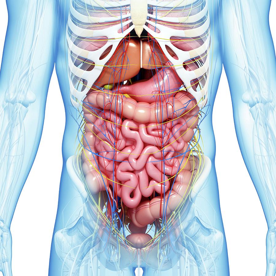
Abdominal Anatomy - Cat Anatomy (Thoracic and Abdominal Organs) : Anatomy of the abdominal wall, inguinal region & hernias, hernias, gut and peritoneal cavity.
Abdominal Anatomy - Cat Anatomy (Thoracic and Abdominal Organs) : Anatomy of the abdominal wall, inguinal region & hernias, hernias, gut and peritoneal cavity.. The anterolateral abdominal wall formed of 4 layer skin, fascia, muscles, and peritoneum. Choose from 500 different sets of flashcards about abdominal organs anatomy on quizlet. Understanding abdominal anatomy and physiology is essential to understanding the human body as a whole. We will wrap up with an overview of several abdominal diseases that might all present themselves with pain. Abdominal cavity, largest hollow space of the body.
Understanding abdominal anatomy and physiology is essential to understanding the human body as a whole. Who better to review abdominal anatomy with, than an experienced expert? A good amount of area is covered by the abdominal wall. Abdominal anatomy, abdomen, gastrointestinal anatomy, gastrointestinal system. 6 write the origin, insertion and nerve supply of muscles of anterior abdominal wall.
Abdominal Anatomy Photograph by Pixologicstudio/science ... from images.fineartamerica.com Lee moffitt cancer center & research institute in. In order to find the right training and to perform the exercises properly, it is important to know what are the abdominal muscles. We will wrap up with an overview of several abdominal diseases that might all present themselves with pain. Anatomy of the abdominal wall, inguinal region & hernias, hernias, gut and peritoneal cavity. • in this module, we will explore basic abdominal anatomy identifiable with common imaging modalities. Identify abdominal anatomical structures in a variety of medical imaging platforms. Introduction to sonographic abdominal anatomy. The above lines intersect and divide the abdomen into nine regions (clockwise from the top)
Compare and contrast the different medical imaging modalities presented in the tutorials.
• in this module, we will explore basic abdominal anatomy identifiable with common imaging modalities. 5 name the nine abdominal regions and their main contents. And inferiorly by the symphysis pubis, pubic tubercle, inguinal ligament, anterior superior iliac spine, and. Two layers in abdomenfatty superficial layer (camper's fascia)deeper membranous layer (scarper's fascia). Lee moffitt cancer center & research institute in. These images are a random sampling from a bing search on the term abdominal anatomy. click on the image (or right click) to open the source website in a new browser window. The linea alba (open arrowhead); Radiology basics of abdominal ct anatomy with annotated coronal images and scrollable axial images to help medical students and junior doctors learning anatomy. A collection of articles covering abdominal anatomy, including abdominal wall anatomy and abdominal cavity anatomy. Simple, easy notes for quick revision of important questions. Become familiar with the anatomical divisions by exploring the world's most advanced 3d anatomy platform in complete anatomy. We created an anatomical atlas of abdominal and pelvic ct which is an interactive tool for studying the conventional anatomy of the normal structures based on a multidetector computed tomography. Anatomical structures of the abdomen and pelvis are visible as interactive labeled images.
Radiology basics of abdominal ct anatomy with annotated coronal images and scrollable axial images to help medical students and junior doctors learning anatomy. Become familiar with the anatomical divisions by exploring the world's most advanced 3d anatomy platform in complete anatomy. In order to find the right training and to perform the exercises properly, it is important to know what are the abdominal muscles. The linea semilunaris (open arrow); Choose from 500 different sets of flashcards about abdominal organs anatomy on quizlet.
Abdominal wall muscles - 3D tutorial - YouTube from i.ytimg.com Identify some abdominal pathology on medical images. Radiology basics of abdominal ct anatomy with annotated coronal images and scrollable axial images to help medical students and junior doctors learning anatomy. Learn about abdominal organs anatomy with free interactive flashcards. Introduction to sonographic abdominal anatomy. The anterolateral abdominal wall formed of 4 layer skin, fascia, muscles, and peritoneum. A good amount of area is covered by the abdominal wall. Windham was previously a surgical oncologist in the sarcoma program of the h. Anatomical structures of the abdomen and pelvis are visible as interactive labeled images.
Anatomical structures of the abdomen and pelvis are visible as interactive labeled images.
Anatomical structures of the abdomen and pelvis are visible as interactive labeled images. We created an anatomical atlas of abdominal and pelvic ct which is an interactive tool for studying the conventional anatomy of the normal structures based on a multidetector computed tomography. A collection of anatomy notes covering the key anatomy concepts that medical students need to learn. The abdominal region is supported by the anterior and posterior abdominal wall that supports the viscera and maintains the posture where there's no bony support. Two layers in abdomenfatty superficial layer (camper's fascia)deeper membranous layer (scarper's fascia). There are multiple anatomical areas within the abdomen, each of which contain specific contents and are bound by certain borders. This muscle forms the anterior and lateral abdominal wall. The abdomen (colloquially called the belly, tummy, midriff or stomach) is the part of the body between the thorax (chest) and pelvis, in humans and in other vertebrates. Sciency root words make anatomical parts harder to memorize. Understanding abdominal anatomy and physiology is essential to understanding the human body as a whole. Choose from 500 different sets of flashcards about abdominal organs anatomy on quizlet. Become familiar with the anatomical divisions by exploring the world's most advanced 3d anatomy platform in complete anatomy. Most students entering ultrasound have some basic understanding of anatomy.
Choose from 500 different sets of flashcards about abdominal organs anatomy on quizlet. And inferiorly by the symphysis pubis, pubic tubercle, inguinal ligament, anterior superior iliac spine, and. Radiology basics of abdominal ct anatomy with annotated coronal images and scrollable axial images to help medical students and junior doctors learning anatomy. The epigastric vessels (long arrow); You will learn the anatomical basis of pain and how to apply this knowledge in the diagnostic process.
Anatomy Of Human Abdominal Vein System Digital Art by ... from fineartamerica.com We created an anatomical atlas of abdominal and pelvic ct which is an interactive tool for studying the conventional anatomy of the normal structures based on a multidetector computed tomography. Abdominal anatomy seen on ct. This muscle forms the anterior and lateral abdominal wall. 6 write the origin, insertion and nerve supply of muscles of anterior abdominal wall. Transversus abdominis muscle internal abdominal oblique muscle rectus abdominis muscle external abdominal oblique muscle pyramidalis muscle. Understanding abdominal anatomy and physiology is essential to understanding the human body as a whole. Compare and contrast the different medical imaging modalities presented in the tutorials. Mr anatomy of the abdominal wall demonstrating the three flat muscles (short arrow);
Introduction to sonographic abdominal anatomy.
The abdominal divisions should be used in conjunction with other diagnostic approaches in order to accurately diagnose a patient's condition. A collection of articles covering abdominal anatomy, including abdominal wall anatomy and abdominal cavity anatomy. Introduction to sonographic abdominal anatomy. The epigastric vessels (long arrow); Abdominal anatomy seen on ct. Compare and contrast the different medical imaging modalities presented in the tutorials. Abdominal cavity, largest hollow space of the body. The abdominal region is supported by the anterior and posterior abdominal wall that supports the viscera and maintains the posture where there's no bony support. The linea alba (open arrowhead); Gsi asked questions about the abdominal membranes to christopher windham, m.d. We created an anatomical atlas of abdominal and pelvic ct which is an interactive tool for studying the conventional anatomy of the normal structures based on a multidetector computed tomography. A collection of anatomy notes covering the key anatomy concepts that medical students need to learn. Identify abdominal anatomical structures in a variety of medical imaging platforms.


0 Komentar